


By Liz Stevens, writer, UV+EB Technology
Advances in technology seem to be benefiting every human endeavor. In healthcare, innovations in additive manufacturing and material engineering are yielding valuable, demonstrable improvements that facilitate better medical training, better medical procedure pre-planning and better medical device design. A university research team in Mexico has focused its efforts on advancing materials science and 3D printing to benefit the surgical arena and has founded a company devoted to commercializing those innovations. Mædditiva, at Tecnologico de Monterrey, Nuevo León, México, has developed technology to produce 3D-printed anatomical models that faithfully represent a patient’s anatomy, that are as materially close as possible to human tissue and that are transparent. The company earned recognition as part of the RadTech Class of ‘24 for its accomplishments.
Surgeons regularly use body part models when planning for or practicing for a complicated surgery. Medical students rely upon models in their studies, and medical device manufacturers need body part models for reference to design their products. Delivering three disparate qualities in one model – a precise representation of an individual patient’s body part, a model with the feel and flexibility of a particular type of human tissue, and a model with high transparency – has been challenging. Engineering a model to be soft and flexible, for example, has called for material choices that do not support the structural strength needed to retain the model’s anatomy. Efforts to make translucent models more transparent – which especially is valuable for vascular models in which blood flow must be visualized – have included sanding and polishing the models, but this calls for a rigid model and the sanding/polishing action alters the model’s anatomical fidelity. Mædditiva has created a methodology to produce anatomically precise, flexible stereolithography-printed models in commercial elastomeric acrylic-based resins with mechanical properties close to those of human aortic tissue, and with a soft thiol-ene coating as a post-processing step to deliver near-transparency. They call their models “phantoms.”
To learn more, UV+EB Technology talked with the company’s CTO and founder, Alan Aguirre-Soto, director, Nanotechnology Graduate Program, PI, Macromolecular and Photo-sciences (MPS) Research Group, and professor, Chemical Engineering in the School of Engineering and Sciences.
Developing the model
To capture the dimensions of a patient’s body parts, Mædditiva uses medical imaging files created through computed tomography (CT) and magnetic resonance imaging (MRI) scans. Once the scans are in hand, a combination of software products is used to process the data for 3D models. “We use a combination of licensed and open-source software,” said Aguirre-Soto, “including Materialise’s Mimics Suite, 3D Slicer and Meshmixer.” These facilitate the segmenting of medical images, visualization, analysis of images, the design of 3D models, model-making project management and navigation for image-guided procedures.
The company has used off-the-shelf VAT photopolymerization machines, including Form2 (SLA) from Formlabs, Form3 (SLA) from Formlabs and Phrozen Mighty 4K (DLP, mSLA) from Phrozen Technology. “For the interior portions of models,” Aguirre-Soto explained, “we mostly have used the Flexible 80A and the Elastic 80A resin from Formlabs. However, we have tested with more rigid commercial resins, such as the Clear Resin, also from Formlabs. We selected commercial resins that are mechanically soft (as much as possible), but that also are listed as ‘clear.’ They are not exactly clear; the base polymer is better described as being translucent. These ‘clear’ materials actually most often are hazy, such that their translucency is not sufficient for use in some medical applications.”
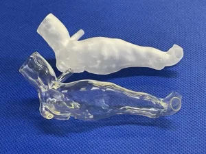
The resin that Mædditiva formulated as a coating truly is clear and applying it via dip-coating mitigates the lack of transparency caused by the relatively rough surface of the printed model (Image 1). “The coating contains thiol-ene monomers,” Aguirre-Soto said. “The idea is that the leftover, unreacted vinyl groups in the surface of the commercial material participate in the thiol-ene click reaction during the curing of the coating. Through this, we achieve a strong-enough addition of the coating to the partially cured base material, as well as allowing the coating to fill the imperfections that led to haziness. Through the latter mechanism, we end up with a coated base polymer material that has a much higher optical transparency.”
Another key feature of the use of thiol-ene chemistry lies in achieving a more uniform polymerization at the model-coat interface and the bulk to prevent the peeling and delamination of the coating. The non-classical chain growth mechanism of thiol-ene chemistry leads to the formation of a more homogeneous network that binds to the 3D-printed object as the bulk coating cures. For the coating, ternary thiol-ene-acrylate chemistry was found to be optimal. A PETMP/allyl glycerol ether/polyethylene glycol diacrylate coating can double the optical transmission of a model from 40% to 80%. The higher transparency correlates with a significant decrease in surface roughness from 2,000 nm to 90 nm after coating. The coating is cured with 365 nm near UV.
Aguirre-Soto described some of the breakthroughs in research and development of the coating formulation. “There were several ‘a-ha!’ moments through the journey toward finding the right coating,” he said. “The thing is that there are a fair number of non-scientific reports in which people mentioned that they tried to coat a printed acrylate object with the same acrylic-based resins. The problem with this approach is that the binding between the coating and the base material is not strong enough. The coating peels.” Another problem, said Aguiree-Soto, is that oxygen inhibition makes it difficult to properly cure the coating on a 3D-printed object. “Furthermore,” he said, “most attempts to coat 3D-printed objects have involved rigid coatings. Here, we wanted to formulate a photopolymerizable coating that was resistant to oxygen inhibition, that would adhere strongly to the base polymer and that formed a transparent and flexible (soft) coat. Once we established these criteria, we had the ‘a-ha!’ moment that this could be achieved with thiol-ene.”
The group’s “phantoms” have been utilized in over 15 complex surgeries and by medical companies to test new stent graft designs. “In our latest use case,” said Aguirre-Soto, “we sent anatomical models to the University of Twente, Netherlands. The models are used in a medical laboratory to gain insight into cardiovascular degenerative diseases.” Mædditiva has a patent for the process of creating high-transparency 3D-printed objects by coating with photocurable resins that react via mechanisms like the thiol-ene click chemistry. The company is in the process of extending its patent coverage internationally.
Additional Notes and URLs:
See the March 2024 Macro-Molecular Rapid Communications published paper on this topic at https://onlinelibrary.wiley.com/doi/full/10.1002/marc.202300611.
Other publications:
Stereolithographic (SLA) 3D Printing for Preprocedural Planning in Endovascular Aortic Repair of a Thoracic Aneurysm at https://journals.sagepub.com/doi/abs/10.1177/15385744231215560
Soft stereolithographic 3D printed phantoms for dual-modality particle image velocimetry (PIV) at https://link.springer.com/article/10.1007/s00348-024-03938-2
For more information, email Aguirre-Soto at alan.aguirre@tec.mx or visit www.linkedin.com/in/alanaguirresoto/.


